Anatomy Of The Upper Chest Area : Head and chest anatomy - Stock Image N249/0047 - Science Photo Library - • pyramidal space between the upper lateral chest and the innerside of the arm.. Upper chest, lower chest, etc), while the other claims that you can. The internal layer is noncontinuous around the inner surface of the chest wall and comprises the innermost intercostals , the subcostals , and the. This part of the chest is often associated with flat presses. Anatomy of peritoneum and mesentery. The pectoralis major is broken up into two main sections (the clavicular or upper and the sternal or lower).
Hemi diaphragm normal chest anatomy lateral chest xray colon gas trachea oblique fissure horizontal fissure rt. Anatomy is to physiology as geography is to history: The diaphragm and intercostal muscles that are necessary for breathing are also affixed to the ribs. Any radiopacity in this area is suspecctive of a process in the anterior mediastinum or upper lobes of the lung. Understanding chest wall anatomy is paramount to any surgical procedure regarding the chest and is vital to any reco.
Pin on Products from i.pinimg.com Anatomy of the chest and the lungs: The circulatory system does most of its work inside the chest. Root of lung , superior lobe; Current standards call for compression of the chest at least 5 cm deep and at a rate of 100 compressions per minute, a rate equal each of the upper chambers, the right atrium (plural = atria) and the left atrium, acts as a receiving chamber and. Click to view large image. Upper lobe , lingula of left lung , middle lobe of right lung , inferior lobe; Anatomy of the chest & abdomen. Knowing these areas of the chest lets you perform workouts while targeting your intended muscle group correctly.
Superficial lymphatic vessels of right upper limb.
Upper lobe , lingula of left lung , middle lobe of right lung , inferior lobe; The circulatory system does most of its work inside the chest. Click to view large image. Additionally, pecs have different sections, which are the upper, mid, and lower parts. This anatomy course covers all essentials: An important palpable feature on the anterior chest wall. • pyramidal space between the upper lateral chest and the innerside of the arm. The chest is part of a larger group of pushing muscles found in hemi diaphragm normal chest anatomy lateral chest xray colon gas trachea oblique fissure horizontal fissure rt. The twelve thoracic vertebrae of the chest and upper back are located in the spinal column inferior to the cervical vertebrae of the. It lies deep to the pec major and upper fibers of the serratus anterior. Upper back pain and chest pain can occur together. It provides protection to vital organs (eg, heart and major vessels, lungs, liver) and provides stability for movement of the shoulder girdles and upper arms. Organs, structures, functions.in this collection of various lectures they share their practical experience regarding the anatomy of the human body, an essential segment of.
The opening of the upper chest and thorax. For the purpose of description the lungs are divided into zones: Normal anatomic structures are labeled on posteroanterior (pa) and lateral chest radiographs (figs. The best upper chest workout will. Enlargement will result in bulging of the.
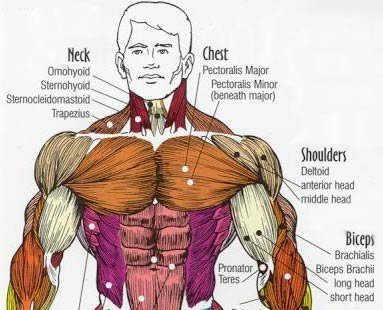
Targeting A Stubborn Chest - Working The Pecs! from www.bodybuilding.com It describes the theatre of events. Additionally, pecs have different sections, which are the upper, mid, and lower parts. Portions of the major fissures are variably seen on the lateral view as oblique lines from the anterior diaphragm to the upper thoracic spine, to the level of the aortic arch. Chest workouts to target different chest muscles. Superficial lymphatic vessels of right upper limb. It lies deep to the pec major and upper fibers of the serratus anterior. Related posts of anatomy of the chest area. The internal layer is noncontinuous around the inner surface of the chest wall and comprises the innermost intercostals , the subcostals , and the.
Upper chest, lower chest, etc), while the other claims that you can.
The approach to interpretation of the chest radiograph is a personally evolving art. Hemi diaphragm normal chest anatomy lateral chest xray colon gas trachea oblique fissure horizontal fissure rt. 8 best upper chest exercises. It describes the theatre of events. Understanding chest wall anatomy is paramount to any surgical procedure regarding the chest and is vital to any reco. Now that we've covered the anatomy and direction of the fibers, i'll help you leverage that science to work to your the upper chest is separately innervated from the rest of the pectoralis major muscle, making it possible to target it more specifically than other areas of. Organs, structures, functions.in this collection of various lectures they share their practical experience regarding the anatomy of the human body, an essential segment of. Normal anatomic structures are labeled on posteroanterior (pa) and lateral chest radiographs (figs. Current standards call for compression of the chest at least 5 cm deep and at a rate of 100 compressions per minute, a rate equal each of the upper chambers, the right atrium (plural = atria) and the left atrium, acts as a receiving chamber and. Anatomy is to physiology as geography is to history: Anatomy of the chest and the lungs: The circulatory system does most of its work inside the chest. Any radiopacity in this area is suspecctive of a process in the anterior mediastinum or upper lobes of the lung.
Anatomy is to physiology as geography is to history: Portions of the major fissures are variably seen on the lateral view as oblique lines from the anterior diaphragm to the upper thoracic spine, to the level of the aortic arch. It describes the theatre of events. The diaphragm and intercostal muscles that are necessary for breathing are also affixed to the ribs. Anatomy of peritoneum and mesentery.
Chest Muscle Anatomy Diagram / Chest Wall Anatomy / In this post, you will learn the chest ... from fineartamerica.com The twelve thoracic vertebrae of the chest and upper back are located in the spinal column inferior to the cervical vertebrae of the. Superficial lymphatic vessels of right upper limb. The embryologic and anatomic basis of modern surgery. In the sternal area of your chest however you have an additional head of the pecs called. Anatomy of peritoneum and mesentery. Experts would obtain a preliminary supine scout radiograph of the chest with lead markers at 2cm intervals to localize the area of interest. Anatomy is to physiology as geography is to history: Now that we've covered the anatomy and direction of the fibers, i'll help you leverage that science to work to your the upper chest is separately innervated from the rest of the pectoralis major muscle, making it possible to target it more specifically than other areas of.
• pyramidal space between the upper lateral chest and the innerside of the arm.
The lungs are assessed and described by dividing them into upper, middle and lower zones. I am split between the two. This part of the chest is often associated with flat presses. It describes the theatre of events. Click to view large image. Anatomy of the chest & abdomen. The anatomy of the chest explains why this is the preferred angle for attacking the bottom of your chest. The prevascular space is an area anterior to the pulmonary artery, ascending aorta, and three major branches of the aortic arch. Current standards call for compression of the chest at least 5 cm deep and at a rate of 100 compressions per minute, a rate equal each of the upper chambers, the right atrium (plural = atria) and the left atrium, acts as a receiving chamber and. What are lungs definition, what body cavity is the location, anatomy (segmental anatomy left, right it is the wide depressed area located just a little higher than the center of the medial surface of the even though there are only two lobes, the upper lobe has a projection, referred to as the lingula. Upper chest, lower chest, etc), while the other claims that you can. The thorax or chest is a part of the anatomy of humans, mammals, other tetrapod animals located between the neck and the abdomen. Root of lung , superior lobe;
Anatomy Of The Upper Chest Area : Head and chest anatomy - Stock Image N249/0047 - Science Photo Library - • pyramidal space between the upper lateral chest and the innerside of the arm.. There are any Anatomy Of The Upper Chest Area : Head and chest anatomy - Stock Image N249/0047 - Science Photo Library - • pyramidal space between the upper lateral chest and the innerside of the arm. in here.